| 
Screening, Diagnosing, and Monitoring Breast Cancer
Focusing on the NO.1 Disease Affecting Women��s Health
Non-invasive: No radiation, no side effect, and no pain
Market Demand:
-One out of ten women will suffer from breast cancer in her life.
-In developed countries, 1/3 of woman cancer patients are breast cancer related
-In China, breast cancer is a dramatically growing disease, and is the No.1disease in urban women population.
-Early detection is a critical step for disease treatment and for improving women's life quality.

Leading Technology:
-OPTIMUS system is developed on the basis of an innovative technology protected by several US and Chinese patents.
-OPTIMUS is the result of a combined research and development effort by the University of Connecticut and MDT since 1998.
-This effort has been supported by a variety of resources including the Connecticut Innovative Fund, NIH grants, Hartford Hospital, and the Health Center of the University of Connecticut.
-As an innovative technology, it has been shown in the media like Laser World Magazine, Radiology Magazine,and featured in television programs on CBS.
Angiogenesis: A common denominator in breast cancer
Abnormal blood supply: An important factor in early diagnosis
Common characteristics of malignant tumor growth: Research has shown solid breast tumors become clinically relevant once they develop a blood supply, and malignant tumors have higher MVD of their surrounding tissues. Subtle vascular changes are often associated with breast cancer in its earliest stages. DOT technology can detect abnormal blood supply changes in local tissues, and thus provide information for early diagnosis before the detection of morphological changes.
OPTIMUS imaging: provides functional information of breast tissues, and reveals the concentration of Hemoglobin and Oxygen saturation, reflecting angiogenic status of lesions.

Diffusion Optical Tomography (DOT):
-Tumor Angiogenesis: In 1994, the Angiogenesis Foundation identified angiogenesis as the ��common denominator�� in important diseases, including breast cancer. Research has shown that solid breast tumors become clinically relevant once they develop a blood supply. Subtle vascular changes are often associated with breast cancer in its earliest stages. Recent technology detects vascular changes and forms a vascular profile to indicate the early vascular zed lesion.
-Detection with Multi-Wavelength �C OPTIMUS employs multi-wavelength DOT scan to measure HBO2 AND Deoxy HB and calculate hemoglobin concentration and tumor hypoxia
-Probe Design �C With unique NIR probes for functional imaging design, OPTIMUS images reflect optical diffusion status in different tissue positions

Dual Modality Imaging:
-OPTIMUS combines the advantages of ultrasound imaging and DOT imaging to provide diagnostic information on breast lesions
-Ultrasound imaging demonstrates structural changes and anatomy of breast tissues, allowing doctors to analyze a multiplicity of conditions and assess lesions
-OPTIMUS imaging provides functional information on breast tissues, and reveals the concentration of hemoglobin and oxygen saturation, which are directly related to angiogenesis of lesion

-Ultrasound Imaging �C Uses the probe in the same way as a regular. ultrasound imaging to locate lesion or areas of interest, and acquire an image
-Laser Scan �C Uses multi-wavelength laser scan to acquire optical information of any suspicious area
-DOT Image Construction �C Reconstructs 3D functional images to reflect the spatial distribution of HBT, HBO2, and HB
-Quantitative Analysis �C Performs quantitative analysis of DOT and ultrasound images to generate a report with BI-Rads rating


Innovative Solution:
-Localization- Uses ultrasound imaging to localize tumors, which offers good spatial resolution, and can detect breast lesions of a few millimeters in size. OPTIMUS takes advantage of this technology to localize tumors.
-Identification- Optical tomography is used to indicate the likelihood of tumor malignancy, a new technique that employs diffused light in the near infrared spectrum, provides functional images of tumor angiogenesis and tumor hypoxia. OPTIMUS utilizes such functional information to indicate the likelihood of tumor malignancy.
-Early Detection- OPTIMUS can detect and diagnose lesions as small as 0.5cm in diameter.
-Non-invasive- OPTIMUS performs breast imaging with non-compression, non-radiation, and non-invasive.
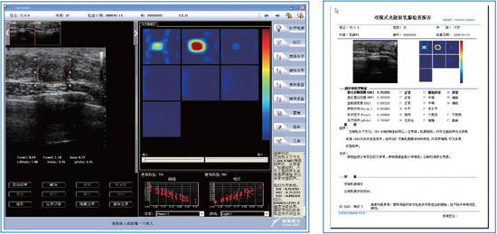
OPTIMAGE-Diagnostic Tools
-Dual modality operation and visualization, simple and easy to use
-Rich information about tumor blood and morphological characteristics, including hemoglobin(HBT), Oxygen concentration(SO2), tumor shape, lobe, and orientation, etc.
-CAD provides standard results in compliance with BI-RADs
-Easy and clear report generation, storage, and PACs compatible

Initial Clinical Trial in US:
HB Index Chart (8 early stage breast cancer, 57 benign tumor)
Clear Efficacy Indication
-The result shows significantly higher HB index values in the malignant group (avg.122µmol/L)than those in the beginning group (avg.55 µmol/L).
-When a threshold of 95 is used to separate malignant and benign lesions, the sensitivity specificity values are 100% and 96%.
-Optical tomography provides higher sensitivity and higher specificity than color doppler ultrasound.

Clinical Trial and Data in China:
HB Index Chart
Clinical Trials
Clinical trials were performed in four hospitals in China. The diagnosis result of OPTIMUS system was compared with X-ray mammography and biopsy results. With 90 trial cases, OPTIMUS system demonstrates 100% sensitivity and 86% specificity.
Screening and Diagnosis
OPTIMUS is used in a hospital specializing in tumor as a screening and a patient diagnosis device. Among 833 women examined over a two-month period, 38 were diagnosed as suspicious malignant lesions. 23 out of the 38 patients underwent surgery with 20 confirmed cancer cases.

|

