SS-2000 color doppler ultrasound system applies advanced digital technology, advanced broadband probe technology, full-featured application software system and a variety of leading-edge image processing technology with delicate image quality and committed system performance to meet clinical requirements.
・Full digital imaging technology ・17 inch LCD monitor ・Up to 4 active probe connectors ・Broadband Multi-frequency probes ・512/1024 frame cine loop ・8 segment TGC ・Powerful information management ・Complete application software packages ・Large capacity hard disk ・USB ports ・Compatible with laser/inkjet printers ・Fashion and ergonomic design ・4D probe and imaging function(optional)
Operating Modes: ・2D mode imaging in fundamental and harmonic mode ・M mode with advanced cine loop and image review ・Color doppler velocity mode ・Power doppler mode(CDE) ・Directional color power doppler mode(DCDE) ・Pulsed wave spectral doppler mode(PW) ・Continuous wave spectral doppler mode(CW) ・Duplex mode ・Triplex mode ・Split screen real time display mode in 2D/2D or 2D/color
User Interface: ・User-centric ergonomic keyboard control panel ・On/Off task light and back-lit illumination of keyboard control panel ・Customized soft key selections displayed on screen provide easy and immediate access control ・Wrist support to help reduce operator repetitive stress injuries ・Multi-directional articulating monitor arm to furtherimprove ergonomics ・Wheel-look mechanism
Extended clinical capabilities: ・Abdominal ・Obstetrical ・Gynecological ・Cardiac ・Pediatric ・Urological and Prostate ・Vascular and small organ ・Neonatal ・Musculoskeletal ・Surgical scanning
――Excellent imaging technology――
Real-time dynamical focusing
It adopts continuous dynamical receiving aperture (CDA) and continuous dynamical receiving focusing (CDF).The Focus of real-time changed when receiving. That’s to say, it can far field gradually according to the depth, so that the ultrasonic beam can be delicate in both short resolution will be greatly improved, so the frame rate will not be lower as the increasing of focuses.
Smoothing processing technology of image
An advanced 2D image automatic filtering device is inserted to the system to realize smoothing processing and improve the clarity of fine structures of the tissues being checked up.
Filtering technique
The filtering device used in this system can filter the noise signal of static and slowly-moving tissues or low-speed blood stream. It’s hard to detect the noise, but this system can improve SNR and improve the quality of image effectively.
――Extraordinary application range――
Endocavity ultrasound
It’s a part of intervention ultrasound, which is mainly composed of vagina, esophagus, recta and gastro scope ultrasound. It can prevent the air and skeleton from affecting ultrasonic examination, and at the same time, it uses high-frequency probe., therefore a clear image can be received.
Intervention ultrasound
It’s branch of modern ultrasonic medicine. Ultrasound puncture technology not only can be used in cytologic examination, histology biopsy, intravenous radiography, and percutaneous puncture intravenous radiography, but also can be used in therapeutic examination including percutaneous puncture drainage technology, medicament injection and so on. It’s simple and convenient, and is characterized by the advantages like high positive rate, safety, sensitivity, and clear image.
3D imaging
The computer can process and rebuild the ultrasonic echo collected according to the required image, and display the three-dimensional image. It’s helpful to locate space, improve spatial resolution, and can make precise quantitative analysis (such as the measurement of volume), so as to provide all-round ultrasound patterns for clinic.
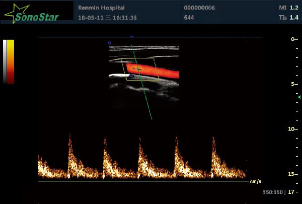
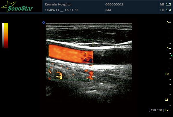
Color Doppler Energy-gram (CDF)
It’s totally new ultrasound pattern processing model. It’s a color blood stream displaying method with Doppler shift strength (amplitude) as the information source, which can get rid of the dependence on detection angle in routine CFM display: meanwhile it won’t generate Alias spurious image, so it can display the minor blood vessels and low speed blood stream clearly.
Color Doppler Direction Energy-gram (DCDE)
It’s a directive Doppler energy-gram, which is the combination of energy Doppler and color Doppler. This technology can combine the advantages of these two. It can display the speed and direction of blood stream, and can display the low speed and low-flow blood stream with Doppler energy-gram as well.
Excellent 2D imagine technology (2D)
It adopts super high density and ultra-wideband probe as well as high-efficiency matching layer and strongly absorbing materials to eliminate near field disturbance for the convenience of observing the shallow tissues. The unique digital signal processing technique can reduce energy loss and increase penetrability to allow the high-frequency probe to detect deep tissues. Besides, it adopts optimization control to physical sign image(PSI) so that patients with different signs can enjoy the same treatment.
Color Doppler Technology (CDT)
It mainly has the ability to provide hemodynamics information, which is called noninvasive angiography clinically. It’s shown as follows:
・It can visually display the distribution and direction of blood stream so as to distinguish the arteries and veins clearly.
・It can help to understand the time phase and velocity of blood stream.
・It can help to find out splitting flow and back-streaming reliably.
・It can conduct quantitative analysis to the origin, width, length and area of blood stream.
Broadband multi-frequency probe technology
Dynamic frequency scanning, a broadband probe technology based on a certain frequency, can realize the simultaneous transmission of multi-frequency and receive high frequency at the near field and low frequency at the far field optionally , thereby to meet the different requirements of clinic. In this way, it can avoid the difficulty and limitation in using high frequency probe(which can lessen greatly in the body) independently in diagnosing the deep organs or foci .
Ultrasound workstation
This system can be connected with DICOM3.O work station directly in windows operating environment. The modular workstation can process image in real time, conduct comp???? digital acquisition , storage, playback and transmission of ultrasound examination, and finis the management of analysis and report,
Application functions of the workstation include:
・Acquisition, storage, playback and transmission of static & dynamic images as well as the management of patient data;
・Perfect integral DICOM3.0 network connection functions, realizing the information-sharing with other section or department office;


 Download the catalog