






Why More Clinics and Hospitals Are Adopting Handheld Ultrasound
- 2026-10-30
- 64
- Guangzhou Sonostar Technologies Co., Limited
Ultrasound used to be defined by where it happened. A patient with a clinical question was sent to an imaging suite, examined by a sonographer, and the answer came back hours or days later. That model has not disappeared, but something has grown alongside it: focused imaging performed by the treating clinician, at the bedside, in the exam room, or in an ambulance, with the answer available in the same conversation.
This is not a marketing story. It is a measurable shift in how imaging is distributed across healthcare facilities, and it is visible in adoption data across both small clinics and large hospital networks.
1. What the adoption data actually shows
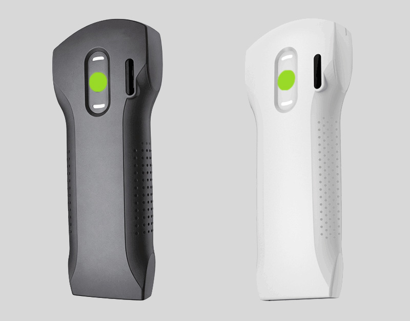
Different research firms size the point-of-care ultrasound (POCUS) market differently depending on whether cart-based systems are included — published estimates for 2025 range from roughly US$2.35 billion to US$6.53 billion for the category. The absolute numbers vary; the direction does not. Every major estimate puts handheld and portable devices as the fastest-growing segment, and every one forecasts continued growth through the next decade.
The more useful figures are penetration rates, because they describe behavior rather than revenue:
Clinic penetration has roughly doubled since 2020. Market research published in mid-2026 estimates that the share of clinics using handheld ultrasound rose from about 22% in 2020 to approximately 38% by mid-2026.
Hospital emergency departments have largely standardized on it. The same research places POCUS integration into formal emergency protocols at roughly 68% of major hospitals in developed markets, with point-of-care echocardiography reported in around 72% of major US hospitals.
It is displacing other imaging, not just supplementing it. Bedside ultrasound has been associated with reductions in CT utilization on the order of 30–35% for certain acute presentations, because the clinical question gets answered before a cross-sectional study is ordered.
Hospitals remain the largest buyer by value (roughly 40–42% of category revenue), but clinics are the segment where new capability is being created rather than upgraded.
Read together, these numbers describe a category that has moved past early adoption. The question for a facility is no longer whether handheld ultrasound works, but how to deploy it sensibly.
2. Five forces behind the shift
Clinical workflow moved to the point of care. Emergency medicine led this transition: focused protocols such as FAST for trauma, lung sliding for pneumothorax, and focused cardiac assessment in undifferentiated shock produced answers fast enough to change management in real time. Once a generation of clinicians trained on those protocols, the expectation traveled with them into primary care, anesthesia, critical care, and outpatient specialties.
The cost structure changed from a capital project to a per-clinician tool. A cart-based system is a capital purchase: a room, a budget cycle, a service contract. A handheld device is closer to a clinical instrument — priced at a level a department or a practice can approve without a facilities project. For a clinic that never had imaging at all, that difference is the entire decision.
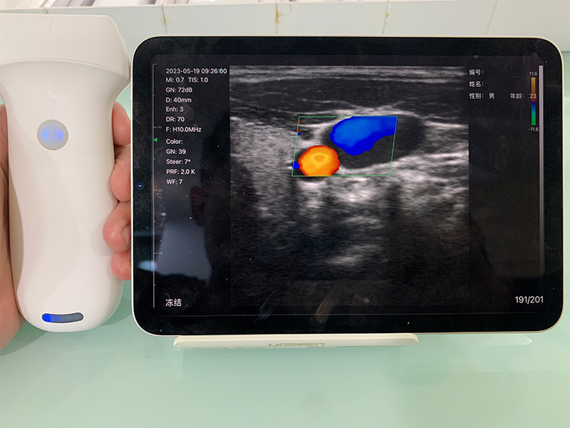
Image quality crossed the diagnostic threshold. Early handheld units were screening tools. Current devices with 64+ channels and 128–192 element arrays produce images that answer defined clinical questions reliably — whether there is free fluid, whether the bladder is full, whether the tendon is intact. That distinction matters more than peak specification: the device does not need to match a premium cart for every study, it needs to be trustworthy for the focused protocols it is deployed for.
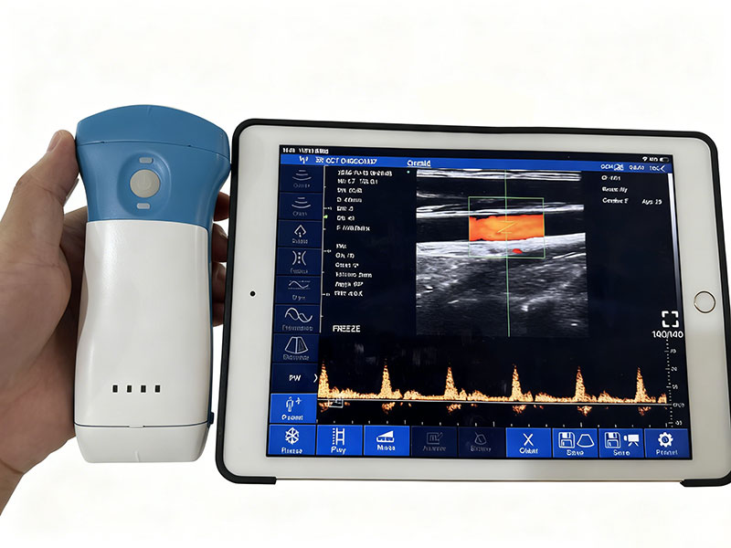
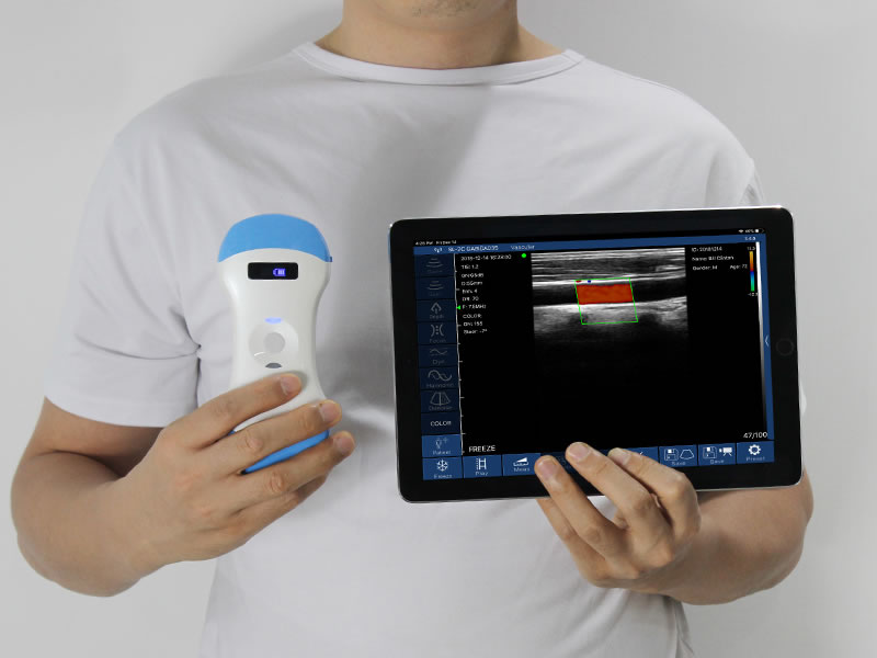
Wireless design removed the console dependency. When the probe itself does beamforming and transmits over Wi-Fi to a phone, tablet, or existing hospital PC, the dedicated display stops being part of the purchase. This is what made the economics work for multi-site deployment — one imaging capability, many rooms, no hardware duplication. Interchangeable and multi-array probes extended this further: a three-in-one convex/linear/phased array probe covers abdominal, vascular, and cardiac scanning in one device, which is what makes a single unit viable across departments.
The training barrier came down. Focused protocols are teachable in days rather than months, and device software now carries presets, measurement automation, and on-screen guidance that reduce dependence on operator experience. Many facilities formalize this through defined protocol sets and quality review rather than open-ended scanning.
3. What adoption looks like in a clinic
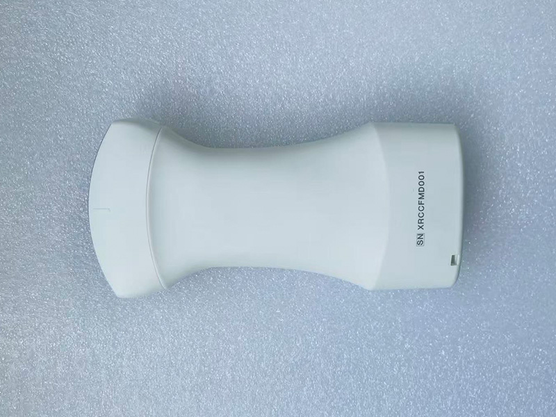
For a primary care or urgent care clinic, the change is categorical rather than incremental. Before handheld ultrasound, the options were to refer out and wait, or to manage without imaging. Neither is satisfying when the question is answerable in ninety seconds.
| Clinical question | Conventional pathway | With handheld ultrasound |
|---|---|---|
| Urinary retention in an elderly patient | Refer to imaging; catheterize empirically and wait for confirmation | Measure bladder volume in the room before deciding to catheterize |
| Soft tissue swelling after injury | Refer or treat empirically | Distinguish fluid collection from solid mass; guide aspiration if needed |
| Difficult peripheral IV access | Repeated blind attempts; escalate to vascular team | Visualize the vein and confirm cannula position |
| Undifferentiated abdominal pain | Refer for imaging; patient returns another day | Targeted screening determines whether referral is urgent |
| Follow-up of a known effusion | Scheduled imaging visit | Assessed during a routine visit |
The economic argument is straightforward: a clinic buys a diagnostic capability it previously could not offer, without adding a room or a staff member. Guidance on matching probe configuration to this kind of mixed caseload is covered in our practical selection guide for medical institutions.
4. What adoption looks like in a hospital
In a hospital, handheld devices do not replace the imaging department — they change where the first look happens. The pattern that has emerged:
Emergency department — screening happens during the primary survey rather than after a radiology request. See our overview of critical care and emergency applications.
Intensive care — daily volume status, pleural assessment, and line placement checks without transporting unstable patients.
Anesthesiology — real-time needle guidance for nerve blocks and vascular access.
Cardiology and internal medicine wards — focused cardiac assessment at the bedside for gross function and effusions.
Vascular and procedural suites — access site evaluation and DVT screening before formal studies.
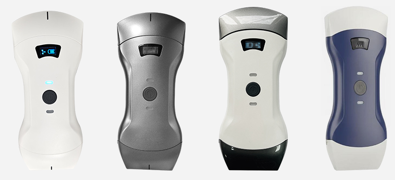
Hospital deployments tend to buy more units but fewer probe types per unit, matching device configuration to department protocol. This is why modular and multi-array probes matter at hospital scale — a convex probe for abdominal work, a linear probe for vascular and superficial imaging, and phased array for cardiac, or a single combined unit where one clinician covers all three.
5. Where handheld ultrasound still falls short
Depth and body habitus. Deep abdominal and pelvic imaging in patients with high body mass index remains challenging. A cart-based system with a lower-frequency probe and more processing headroom still performs better.
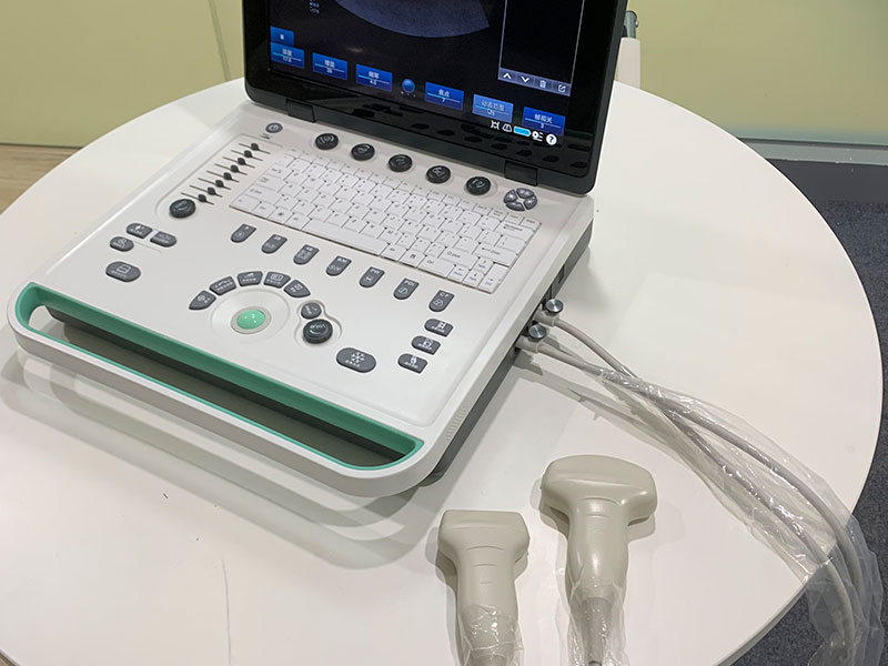
Comprehensive studies. Full echocardiography with Doppler quantification, detailed fetal anatomy surveys, and complete vascular duplex studies remain cart-based work.
Operator dependence. The device does not interpret the image. Outcomes correlate with training, which is why protocol definition and periodic quality review matter more than hardware specification after the first month.
Documentation and workflow integration. Images that are not saved, coded, and linked to the record create clinical and medicolegal gaps. Ask about DICOM export, PACS/EMR integration, and image archival before purchase — this is routinely the least-planned part of a rollout and the most common source of regret.
Durability and service. Devices that travel between rooms get dropped. Enquire about drop ratings, ingress protection, battery replacement, and turnaround on repair.
6. Is it right for your setting?
The honest answer depends on what you already have:
Small clinic or practice with no imaging today — highest marginal value. One device adds an entire diagnostic category. Start with a probe configuration matching your most common presentations, define two or three protocols, and expand from there.
Urgent care or multi-site outpatient group — strong fit, but standardize. Pick one device, one protocol set, and one training path across sites; inconsistent deployment is what turns a good tool into an unused one.
Hospital department — expect an efficiency gain, not a new capability. Budget for training time and image archival integration, and define which questions the handheld answers versus which still go to radiology.
Dedicated imaging department — handheld units are an adjunct for procedural guidance and portable overflow, not a primary system.
Before you buy: the questions that actually predict satisfaction
Which three clinical questions will this device answer in our setting? If the answer is unclear, deployment will stall.
Is there an annual software subscription, and what happens to functionality if it lapses? Compare total cost over three to five years, not the purchase price.
Can images be exported in DICOM and attached to our existing record system?
Is the battery user-replaceable, and is a spare available?
What are the warranty terms, repair turnaround, and availability of loaner units?
What regulatory clearances apply in our market — FDA 510(k), CE under MDR, or local registration?
Mapping this to your facility
SonoStar has manufactured wireless probe-type handheld ultrasound since 2011 and ships to more than 100 countries. If you are evaluating deployment — whether a single clinic device or a multi-department rollout — our applications team can help match probe configuration to your actual case mix.
Browse handheld ultrasound systems · Request specifications and a quotation · OEM and customization
References
Value Market Research, Global Point-of-Care Ultrasound System (POCUS) Market, 2026 — market size and handheld/portable share.
DataM Intelligence, Global Point-of-Care Ultrasound Market, 2026 — segment growth and handheld share of category.
MarketIntelo, Handheld Ultrasound Devices Market, June 2026 — clinic penetration, hospital protocol integration, and ED utilization estimates.
Precision Reports, Point-of-Care Medical Imaging Market, 2026 — emergency department and critical care adoption indicators.
Publicly announced regulatory milestones from GE HealthCare, Philips, Butterfly Network, and Clarius (2025–2026) — category maturity indicators.
- Pre:Handheld Ultrasound Comparison: Wireless Probe Brands Side by Side
- Next:No more

 Home
 Product
 News
 Contact