






POCUS in Primary Care and Critical Care: What Changes
- 2026-10-28
- 56
- Guangzhou Sonostar Technologies Co., Limited
"Point-of-care ultrasound" reads as one thing, but in practice it describes two activities with almost nothing in common besides the hardware. In a primary care clinic, POCUS answers a question that would otherwise take three weeks and a referral. In an intensive care unit, it answers a question where the acceptable delay is measured in minutes. Same machine, different tolerance for failure. Understanding that difference is what separates a device that gets used daily from one that ends up in a cupboard — and it is the single most useful thing to establish before writing a specification. If you are new to the concept entirely, start with the handheld ultrasound buyer's guide, then come back to this comparison.
What POCUS actually means
POCUS is a focused ultrasound examination performed and interpreted by the treating clinician, at the patient's side, to answer one specific question. That last clause is what separates it from conventional sonography. A radiology study documents everything the protocol covers; a POCUS exam asks "is there free fluid," "is this vessel suitable for a line," or "is the bladder distended," and stops once that question has an answer.
Two consequences follow, and they explain the whole rest of this article:
It is clinician-operated, not department-operated. The person scanning is the person making the decision, so the device has to fit their workflow rather than a sonographer's.
It is question-driven, so the question determines the probe. Settings ask different questions; therefore settings need different arrays.
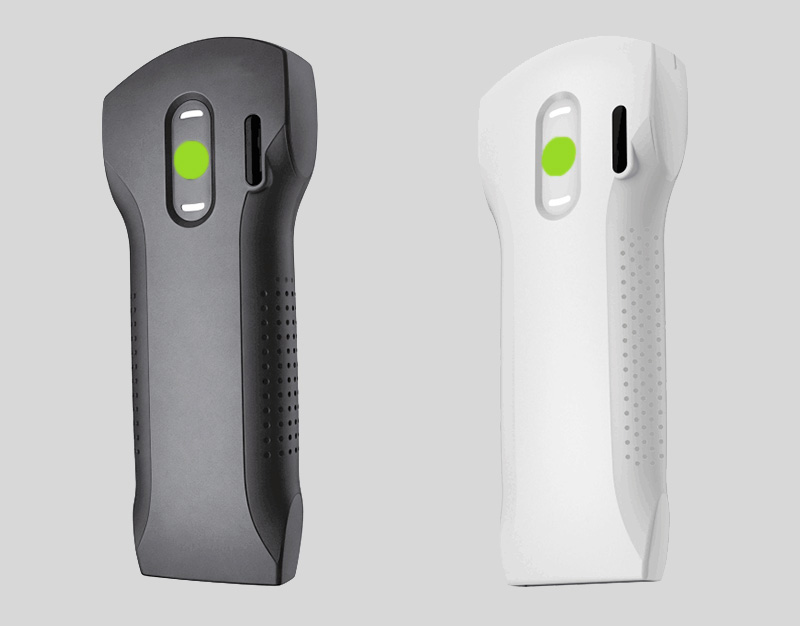
Primary care: answering questions that would otherwise wait
In a clinic or general practice setting, the dominant constraint is the consultation slot. Whatever the clinician does has to happen inside roughly ten to fifteen minutes, alongside everything else in the visit.
Common uses reported across primary care settings include:
Focal abdominal assessment — evaluating a specific area of tenderness, assessing for aortic aneurysm in at-risk patients, or observing gallbladder findings in a patient with compatible symptoms.
Urinary questions — estimating bladder volume and post-void residual in retention, recurrent UTI, or new incontinence.
Vascular access — assessing vein calibre and patency before cannulation in patients with difficult access.
Superficial lumps — characterising whether a palpable finding is cystic or solid before deciding on referral urgency.
Musculoskeletal and injection guidance — visualising a joint or tendon before a therapeutic injection.
The pattern across all of these is that they replace a delay, not a department. A referral that would take weeks becomes a decision made during the visit. That is where the value sits, and it shapes what the equipment must do: it has to be reachable and ready within the consultation, which favours a device that is already in the room and boots quickly.
Array requirements follow the questions: convex for abdominal views, linear for superficial structures and access. Because primary care sees a wide mix rather than one presenting complaint, broad coverage generally beats deep specialisation. Detail: types of ultrasound probes.
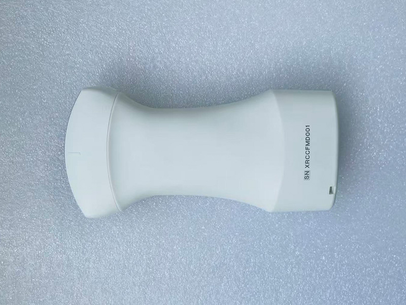
Critical care: minutes, not appointments
Critical care inverts the constraints. There is no consultation slot to protect because there is no queue — but the decision window is far shorter, and the consequences of a missed finding are far larger.
Typical critical care applications include:
Focused assessment in undifferentiated shock or breathlessness — integrating heart, lung, inferior vena cava, and abdominal views to narrow a differential at the bedside.
Pericardial assessment — observing for pericardial effusion when tamponade is in the differential.
Lung assessment — evaluating pleural sliding and B-lines as part of a dyspnoea or fluid-tolerance assessment.
Volume status — assessing intravascular volume and responsiveness as one input among many.
Procedural guidance — central venous access, arterial line placement, thoracentesis, and paracentesis under direct visualisation.
Here, equipment requirements harden considerably:
Phased array is effectively mandatory. Cardiac windows require a small footprint that fits between ribs. Real-world details are covered in our phased array cardiac POCUS guide.
Spectral (PW) Doppler is not optional. Any flow or valvular assessment depends on it.
Time to first image matters. In an arrest or a deterioration, thirty seconds of app-loading delays care.
Failure is expensive. A device that drops its wireless connection mid-procedure is not merely inconvenient.
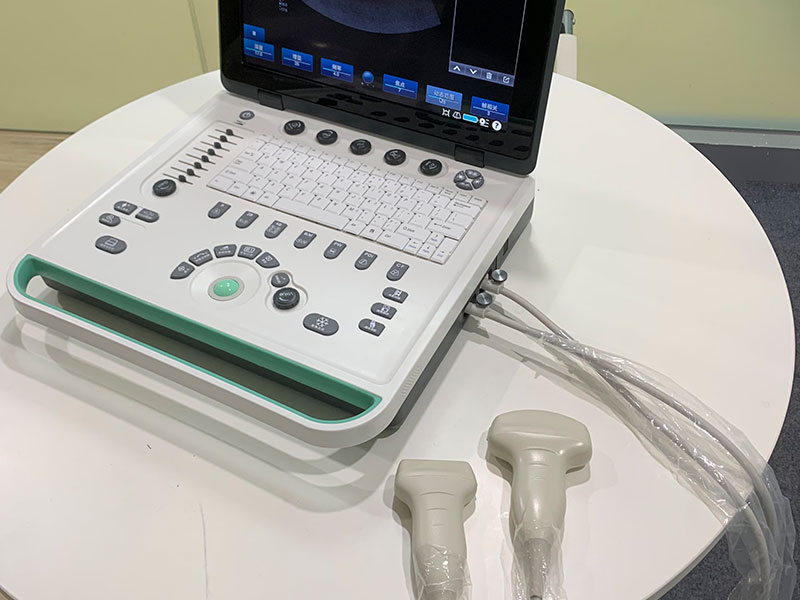
Those requirements are why some units that happily use wireless probes in clinic keep a cart-based system available for critical care — not because wireless imaging is inadequate, but because the risk profile is different. See the honest comparison in laptop vs handheld vs cart.
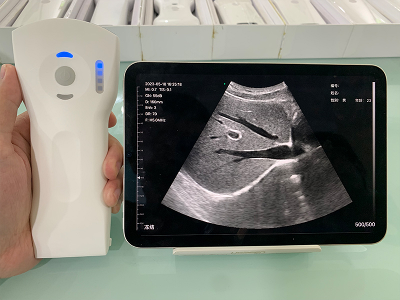
Six differences that change what you should buy
| Dimension | Primary care | Critical care |
|---|---|---|
| Decision window | Days to weeks (replacing a referral) | Minutes |
| Question type | Broad mix across body regions | Concentrated cardio-thoracic and procedural |
| Core arrays | Convex + linear; phased is a bonus | Phased + linear essential; convex for FAST |
| Doppler | Colour useful; PW rarely required | Spectral PW required |
| Tolerance for setup friction | Moderate — within a consultation | Very low — seconds count |
| Training depth expected | Focused, protocol-based competencies | Broader, with heavier emphasis on integration |
Read down the Doppler row and you will see the fork most buyers miss. A device specified for clinical breadth will satisfy primary care completely and fail a cardiac requirement. If one device has to serve both settings, it must be specified against the harder setting, not the easier one.
Specifying equipment for each setting
A primary care configuration
Arrays: convex and linear at minimum. A 3-in-1 adds cardiac and lung capability cheaply enough that it is usually the better first buy — see the 3-in-1 wireless probe guide.
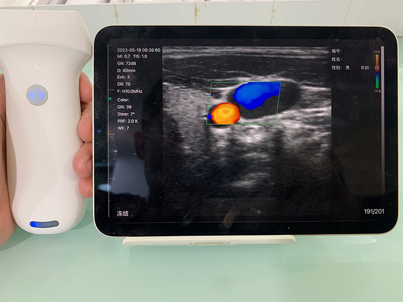
Doppler: colour Doppler for flow and vessel localisation. Spectral PW is rarely needed.
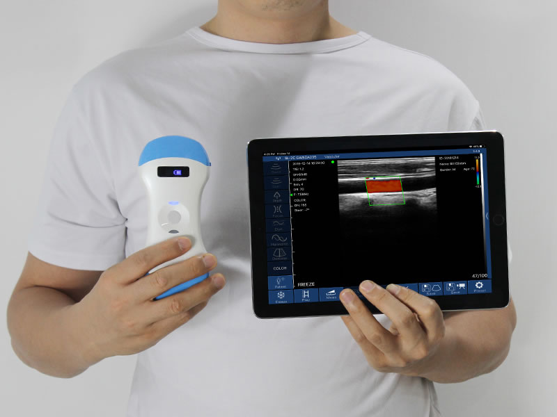
Display: a tablet is often more comfortable to scan with than a phone for abdominal work, and easier to show a patient.
Session length: short intermittent scans, so battery is rarely the constraint.
A critical care configuration
Arrays: phased array with spectral Doppler is the non-negotiable core. Add linear for vascular access and convex for abdominal views.
Readiness: the device should be on, paired, and scanning within seconds. Evaluate time-to-image during a trial, not from a datasheet.
Connection stability: test wireless behaviour under real ICU conditions — many devices on the 2.4 GHz band, metal, and shielding.
Documentation: confirm how images and clips reach the patient record before buying, not after.
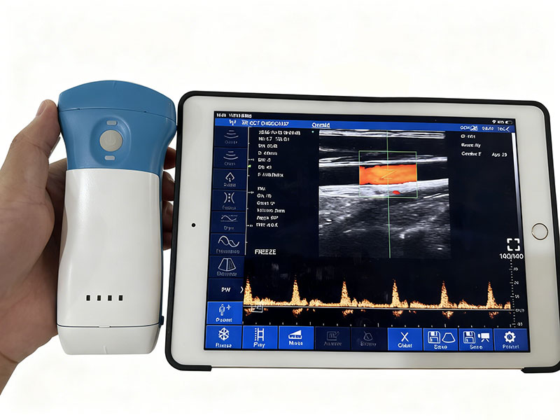
Products that map to these needs: 5PA phased array probe, 8L linear probe for access work, and dedicated units in the wireless probe range. Procurement and pricing detail sits in how much a handheld ultrasound costs.
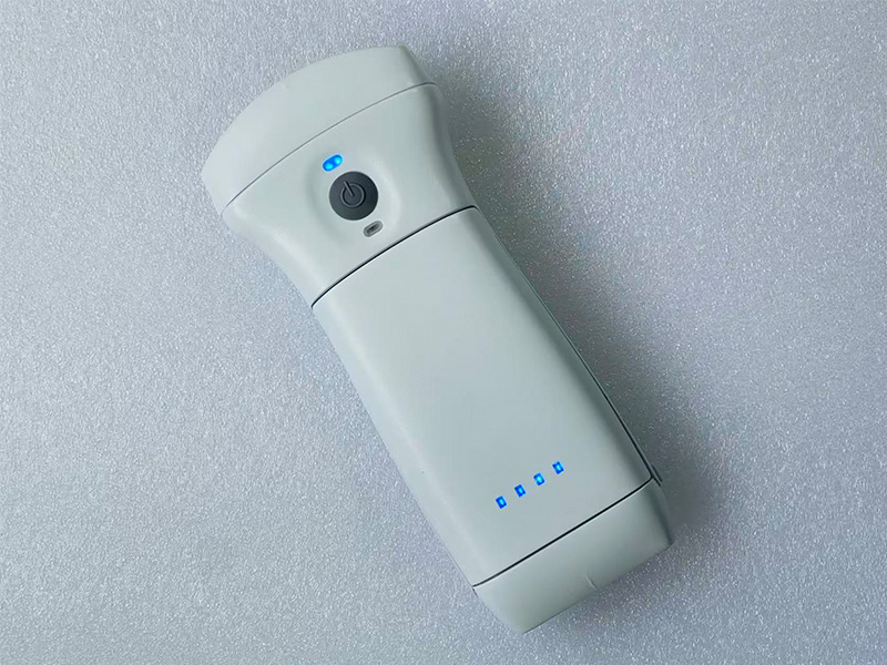
Training and governance differ just as much
Buyers budget for hardware and forget that the same device carries different governance obligations in different hands.
Competency frameworks. Primary care often uses protocol-driven competencies — a defined set of acquisitions a clinician is signed off on. Critical care programmes tend to be broader and more integrated with resuscitation and procedural training.
Supervised practice. Both settings require logged, reviewed scans. Neither is satisfied by a half-day workshop.
Image retention. Whatever is acquired should be stored and reviewable, both for patient care and for quality assurance. Confirm storage and export before purchase.
Scope of practice. What a clinician may perform and act upon is governed locally. That is a policy question, not a device question — and it should be settled before a device arrives.
If you are building a programme from the beginning, start with how to use a handheld ultrasound: a POCUS beginner's guide, and see handheld ultrasound in emergency medicine for a third setting with its own constraints.
Mistakes worth avoiding
Specifying for the easy setting. Buying to satisfy clinic workflows and then expecting ICU performance. Always specify against the harder use case.
Assuming "Doppler" means spectral Doppler. Colour Doppler and PW are different capabilities. Ask which one is included.
Skipping the infection-control question. In high-throughput environments, whether a probe can be immersed or wiped matters daily. Check the stated ingress rating.
Treating training as optional. The pattern behind most unused devices is not technical failure — it is that nobody felt confident reading the images.
Over-buying for a narrow panel. A clinic that scans two body regions does not need the most capable device available; it needs the right two arrays at a sensible specification.
Where SonoStar fits
Stated plainly so you can weigh it against everything above:
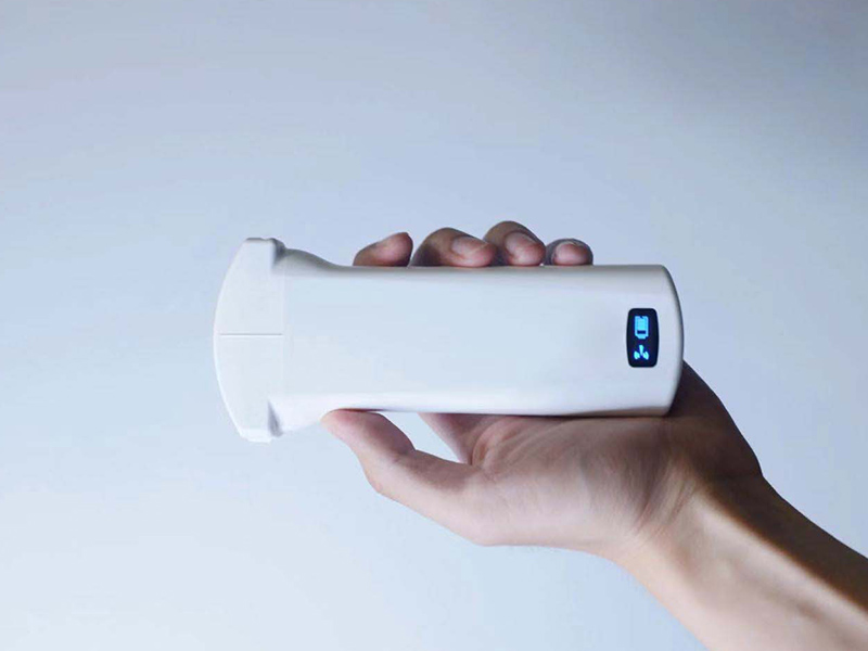
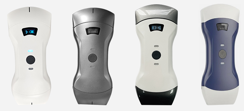
Broad array coverage in one housing. Convex, linear, and phased combinations suit mixed primary care lists, with dedicated single-array probes where volume justifies them.
Full Doppler suite. Colour, power, and spectral PW modes available — the specification that determines whether a device can serve critical care.
Certifications on file. FDA clearance K211321, CE marking (CE 2862), ISO 13485.
Manufacturer-direct. No distributor margin between factory and clinic, which keeps equivalent specifications lower in their price band.
The honest limitation. A smaller brand ecosystem than the largest global names. Institutions building standardised programmes with central credentialing and QA infrastructure may find the large ecosystems worth their premium. For a clinic or a unit buying capability rather than infrastructure, that premium is often hard to justify.
Match the device to the setting
Convex, linear, phased, and 3-in-1 wireless probes — tell us your scan mix and we will specify against the harder use case.
Frequently asked questions
What is POCUS?
A focused ultrasound examination performed and interpreted by the treating clinician at the patient's side, to answer one specific clinical question rather than to produce a comprehensive imaging study.
How is POCUS used in primary care?
Commonly to assess focal abdominal complaints, evaluate bladder volume and post-void residual, assist vascular access, characterise superficial lumps, and guide joint or soft-tissue injection — bringing the answer into the consultation rather than weeks later.
How is POCUS used in critical care?
For focused assessment in undifferentiated shock or breathlessness, pericardial assessment, lung assessment, volume status evaluation, and real-time procedural guidance. The decision window is minutes.
Which probe does each setting need?
Primary care needs convex and linear, with phased array useful but optional. Critical care needs a phased array with spectral Doppler, plus linear for access and convex for abdominal views.
Can one device serve both a clinic and an ICU?
Often yes, particularly a 3-in-1 wireless probe with a full Doppler suite. The compromise to verify is cardiac performance and time-to-image — specify against the harder setting, not the easier one.
Does POCUS require training?
Yes. Acquisition is learnable relatively quickly; interpretation and knowing the limits of a focused exam require structured training and supervised practice. Most professional bodies treat POCUS as a credentialed competency.
How is POCUS used across other departments?
Emergency medicine, anaesthesia, musculoskeletal medicine, obstetrics, urology, and veterinary practice each impose their own constraints. There is a full breakdown in which departments use portable ultrasound most.
This article is provided for general educational and procurement-planning purposes. It is not medical advice and does not constitute clinical guidance or a recommendation to perform any examination. Point-of-care ultrasound must be performed and interpreted by appropriately trained clinicians working within their local scope of practice and regulatory framework; a focused bedside examination does not replace comprehensive imaging or specialist interpretation where indicated. Device specifications reflect manufacturer product information reviewed in 2026 and are subject to change — confirm current specifications, certifications, and regulatory clearance for your market with the manufacturer before purchase.
- Pre:Handheld Ultrasound: The Complete Buyer's Guide (2026)
- Next:No more

 Home
 Product
 News
 Contact