






Are Ultrasound Stethoscopes Accurate? Breaking Reliability into Three Hardware Layers
- 2026-09-10
- 891
- Guangzhou Sonostar Technologies Co., Limited
"Is it accurate?" is the first question buyers ask about an ultrasound stethoscope, and the least useful answer is a blanket yes. Reliability is not a single property. It splits into three layers: spatial resolution (can the device separate adjacent structures), measurement fidelity (does the on-screen value match the real anatomy), and operator consistency (does the same patient scanned twice yield the same answer). A Chinese group standard, the T/CSBME Technical Specifications and Applications of Handheld Ultrasound (Ultrasonic Scope), formalizes exactly this distinction—and its drafting organizations include the Guangzhou-based manufacturer SonoStar alongside the Third Medical Center of the PLA General Hospital, the Shenzhen Institute of Advanced Technology and Tongji Hospital. This article uses that standard and published probe specifications to show how each reliability layer can be checked.
1. How the Standard Defines an Ultrasound Stethoscope
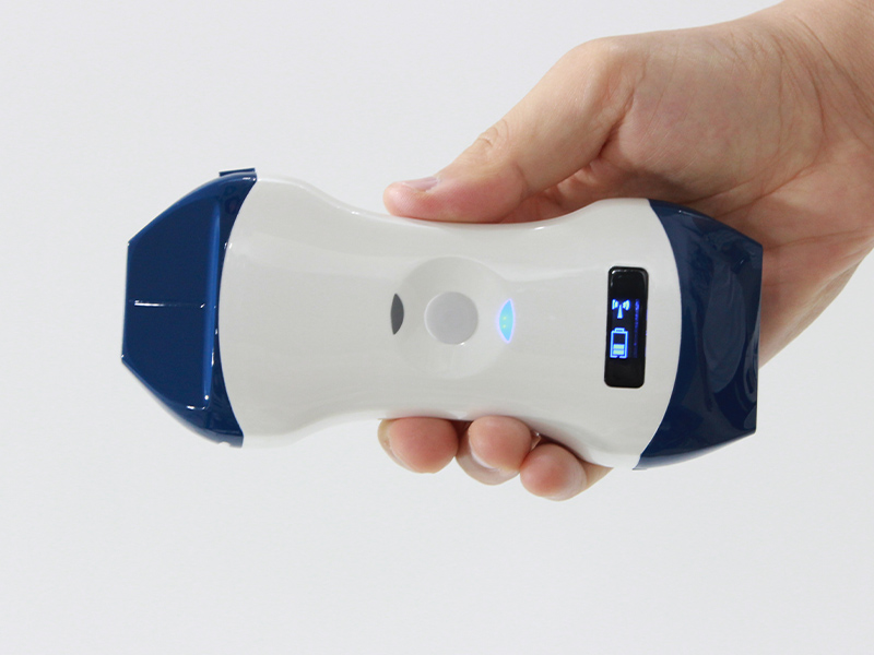
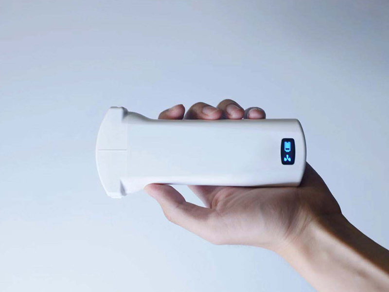
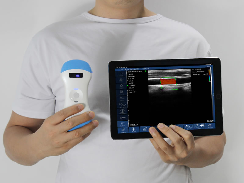
The T/CSBME standard, proposed by the Chinese Society of Biomedical Engineering, gives the category a formal term—ultrasound optoscope, also called handheld, pocket or visual ultrasound—and describes it as the clinician's "third eye," following inspection, palpation, percussion and auscultation. Its physical definition is specific: smartphone-sized, weighing 80–300 g, battery runtime of 2–8 hours, operating temperature −5 °C to 40 °C. Devices are classified as wireless (probe streams to a separate screen over internal Wi-Fi), wired, or all-in-one.
The standard also draws the role boundary explicitly. An ultrasound stethoscope is intended for rapid examination and preliminary screening by clinicians who are not imaging specialists—bedside rounds, pre-hospital assessment, guided puncture—not for replacing a comprehensive diagnostic ultrasound study. That boundary is the starting point for any accuracy discussion: the device is engineered to answer focused questions dependably, not to reproduce a full cart-based examination.
2. Layer One — Spatial Resolution: Elements and Channels
The first layer asks a concrete question: at the target depth, can two nearby structures be told apart? Two hardware numbers answer it.
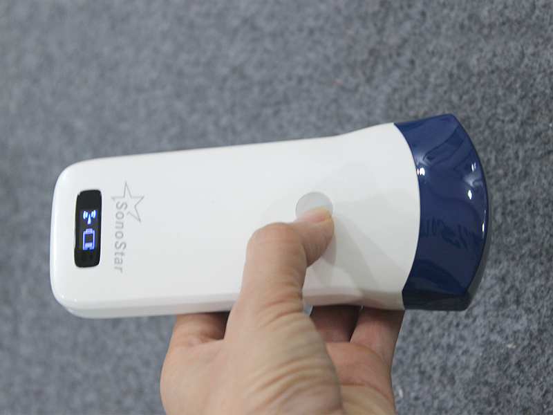
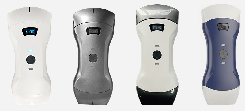
Elements are the piezoelectric pieces in the probe face; their count sets the spatial sampling density of the beam. Channels are the independent transmit/receive electronic paths behind them; their count sets beamforming precision, focusing and side-lobe suppression. The two must be read as a pair. SonoStar's generation difference illustrates the point: the older 3C convex probe carries 80 elements driven by 16 channels, while the current 9CPL three-in-one probe carries 192 elements with 64 channels, and the 6X linear puncture-guidance probe carries 192 elements with 32. A 192-element array multiplexed through 16 channels cannot exploit its own aperture; 192 paired with 64 can.
Frequency then sets the resolution-depth boundary. The 9CPL sheet lists convex 3.2/5.0 MHz with depths to 305 mm, linear 7.5/10.0 MHz at 20–80 mm, and phased-array 3.2/5.0 MHz for cardiac windows. High frequency resolves finer detail but attenuates faster; no processing algorithm reverses that physics. A device whose frequency band does not reach the depth or detail a department needs will be "inaccurate" for that department regardless of brand.
3. Layer Two — Measurement Fidelity: Processing Chain and Image Quality Control
Raw echoes are not yet an image, and the second reliability layer depends on how the processing chain is tuned. Published adjustable ranges on current wireless convex probes include total gain of roughly 30–105 dB, dynamic range in steps from 40 to 110, five-level noise reduction (0–4) and pulse-inversion harmonic imaging. Each control repairs a specific defect: gain rescues an anechoic deep field or suppresses a noisy one; wide dynamic range preserves grayscale gradation in solid organs; harmonic imaging reduces near-field clutter through difficult acoustic windows.
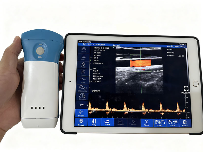
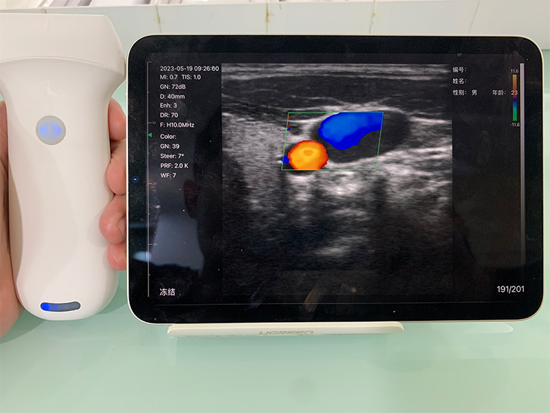
The T/CSBME standard turns this layer into three explicit image quality-control criteria: grayscale images must display skin and subcutaneous layers clearly with the examined area easy to interpret; color Doppler must show vessels well filled, without color bleed-out or signal loss; spectral Doppler envelopes must be complete and sharp enough to read peripheral vessels. A pocket scanner whose auto mode passes these three tests on average patients but locks the operator out of manual adjustment will fail them on difficult patients—and the measurement values derived from those images will fail with them.
The wireless display path belongs to this layer too. Current probes transmit over internal Wi-Fi at around 10 frames per second without needing an external network, but the phone or tablet serving as the screen also affects brightness, latency and perceived sharpness; the display terminal is part of the imaging chain and should be standardized.
4. Layer Three — Operator Consistency: The Layer No Spec Sheet Fixes
The third layer is human, and the T/CSBME standard does not leave it implicit. It requires users to be medical workers trained in basic ultrasound physics and scanning skills, regardless of whether they come from an imaging department.
The same probe produces different results in different hands. A probe placed outside the correct acoustic window, a gain setting pushed to an extreme, or a measurement caliper traced along the wrong wall all manufacture confident-looking errors. This is not a theoretical concern: in the study discussed in the 2003 Heart editorial "The 'ultrasonic stethoscope': is it of clinical value?", roughly one in five patients examined with a pocket imager still required a subsequent detailed echocardiographic examination. A written focused-scanning protocol and supervised practice are therefore components of accuracy, not optional extras.
5. When a Pocket Device Is Simply Not Accurate Enough
Four ceilings are physical, not marketing concessions. First, an ~200 g sealed probe houses fewer channels and a smaller aperture than a cart, so weak Doppler sensitivity, deep penetration and grayscale detail remain lower; comprehensive quantitative diagnosis still belongs on a high-channel system. Second, a fanless sealed housing must manage heat and battery, capping sustained frame rate and transmit voltage. Third, around 10 frames per second supports focused visual assessment, not continuous high-frame-rate quantitative cardiac analysis. Fourth, the standard itself separates documentation tracks—a brief visual-exam record versus a full diagnostic ultrasound report—and blurring that distinction overstates what the device is certified to do.
6. How to Verify Accuracy Claims Before Purchase
Read elements and channels together rather than as isolated figures; confirm the frequency bands actually cover the depths the department scans; verify electrical safety compliance with the IEC 60601-series equivalents of China's GB 9706.1 and GB 9706.237 cited in the standard, plus the standard's requirement of normal operation after three continuous scanning hours; and finally, test demonstrators on the department's own difficult-body-habit patients rather than on factory phantoms, while asking what training protocol the supplier provides.
Conclusion
An ultrasound stethoscope is neither inherently accurate nor inherently unreliable. Its accuracy is the stacked result of element-and-channel architecture, a tunable processing chain meeting defined image quality-control criteria, and a trained operator following a focused protocol. The T/CSBME standard's 80–300 g, 2–8-hour physical definition describes the tool; the three-layer test describes how to judge whether a specific unit's results deserve to be trusted.
Disclaimer: This article is for medical device technology education only and does not constitute clinical diagnostic or procurement guidance. Group-standard text is cited for technical reference; device specifications are subject to manufacturers' official documentation, and clinical use must follow institutional protocols and applicable regulations.
References
[1] Chinese Society of Biomedical Engineering. Technical Specifications and Applications of Handheld Ultrasound (Ultrasonic Scope) (T/CSBME group standard), National Group Standard Information Platform, 2023.
[2] Salustri A, Trambaiolo P. The "ultrasonic stethoscope": is it of clinical value? Heart. 2003;89(7):704–706. doi:10.1136/heart.89.7.704
[3] EFSUMB Position Paper on the Use of Handheld Ultrasound Devices in Clinical Practice. Ultraschall in der Medizin / European Journal of Ultrasound, 2019.
[4] Szabo TL. Diagnostic Ultrasound Imaging: Inside Out. 2nd ed. Academic Press (Elsevier), 2014.
[5] SonoStar official technical specifications: 9CPL, 6X and 3C wireless probe series.

 Home
Home Product
 News
News Contact